Crohn’s disease (CD) is a chronic inflammatory disease of the intestine, of unknown etiology, which is characterized by a discontinuous, segmental manifestation and implication of all intestinal layers. Extra-intestinal manifestations of CD, characterized by inflammatory conditions outside the digestive tract, occur in about 25% of patients.1 Peripheral arthritis, erythema nodosum, pyoderma gangrene, episcleritis, anterior uveitis, oral recurrent ulcer, and ankylosing spondylitis are the most common extra-intestinal manifestations.2 However, inflammatory bowel disease (IBD)-related lung diseases are increasingly recognized. IBD-related respiratory diseases have a variety of clinical manifestations, which can involve the airway and lung interstitial pulmonary vessels, and usually appear several years preceding the diagnosis of IBD.3,4 Ulcerative colitis (UC) is more likely to involve the respiratory system than CD, and lung involvement can aggravate the condition of IBD and is a risk factor for poor prognosis.5
The clinical symptoms of IBD-associated lung disease are hidden, which increases the difficulty of diagnosis. Zhao et al6 found that only 2.7% of IBD patients had respiratory symptoms. Karadag et al7 reported that 15 UC patients were complicated with ground-glass changes in the lungs, but none of them felt uncomfortable. During the time of Corona virus disease (COVID-19), the imaging signs of novel coronavirus pneumonia can also present as interstitial lung disease (ILD), which is difficult to distinguish from the extraintestinal manifestations of CD.
We present a case of a patient with multiple intestinal ulcers associated with ILD in the time of COVID-19. This case had no pulmonary symptoms, which was consistent with previous studies. The purpose of this article is to elaborate on these rare extraintestinal manifestations of CD and to reveal that IBD-related ILD responds excellently to systemic steroid therapy. Moreover, the importance of short-term follow-up chest computed tomography (CT) for differential diagnosis is emphasized.
Case Presentation
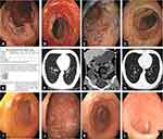
On December 9, 2020, a 27-year-old male patient, who is a student, presented with chief complaints of “loose stools for 4 months, intermittent low-grade fever for 1 month”, and he was admitted to the Departments of Gastroenterology, The First Affiliated Hospital of Wannan Medical College. His complaints did not include abdominal pain, abdominal distension, bloody purulent stool, cough, expectoration, chest tightness and shortness of breath. And a physical examination revealed no abnormalities. This patient has just returned from Japan (November 2, 2020) and has been quarantined for 14 days twice. In Japan, colonoscopy (October 7, 2020) showed multiple ulcers between the sigmoid colon to the terminal ileum (Figure 1A–D), and pathology found that inflammatory cell infiltration in intestinal mucosa and epithelioid granuloma in submucosa (Figure 1E). Regular use of mesalazine did not significantly improve the symptoms of loose stools. Meanwhile, taking levofloxacin orally was not effective for his fever.
Figure 1 (A–D) Colonoscopy showed segmental intestinal disease; (E) pathology found inflammatory cell infiltration in intestinal mucosa, epithelioid granuloma in submucosa; (F) chest CT scan showed multiple patchy ground-glass shadows; (G) a total gastrointestinal CT angiography showed segmental leaping thickening in part of colon; (H) a repeat chest CT scan after systemic steroid therapy; (I–L) a repeat colonoscopy after three infliximab treatments.
Laboratory investigations revealed a normocytic anaemia (hemoglobin 92g/L), hypoproteinemia (albumin 28.8g/L), raised inflammatory markers (C-reactive protein (CRP) 72.9mg/L, erythrocyte sedimentation rate (ESR) 74mm/h) and thrombocytosis (PLT 675*10^9/L). Examination of pathogenic microorganisms suggested that rubella virus immunoglobulin G (IgG) was 208.5IU/mL and cytomegalovirus IgG was 184.4U/mL, which were exceeds the normal limit. Fecal bacteria proportion showed that gram-positive coccus 10%, gram-positive bacillus 10%, gram-negative bacillus 80%, and no fungal spores were found. Fecal occult blood (OB) is negative, and no obvious abnormality was observed in tuberculosis antibody (TB-AB), tuberculous infection with T cells (T-sport), purified protein-derived tuberculin (PPD), blood coagulation, routine before blood transfusion and tumor markers.
The next day (December 10, 2020), the patient developed a high fever with a body temperature of 39.8°C. A chest high resolution computed tomography (HRCT) was urgently performed and result showed infectious lesions in the upper lobe of the right lung and the lower lobe of both lungs, with multiple patchy ground-glass shadows and nodular ground-glass shadows, which suggested interstitial pneumonia (Figure 1F). Then, multiple novel coronavirus (2019-nCoV) pneumonia nucleic acid test was performed, and the results were all negative. Despite the empirical antibiotic and antiviral therapy that was started, his fever remained.
On the third day after admission (December 12, 2020), laboratory investigations revealed that fecal OB was positive and hemoglobin dropped from 92g/L to 84g/L. On the other hand, fecal general flora, Salmonella and Shigella culture were all negative. Acid-fast bacilli and general bacterial cultures in sputum were also negative. Gastroscopy showed chronic superficial gastritis. A total gastrointestinal CT angiography showed that segmental leaping thickening in part of jejunum, distal ileum, ileocecal, ascending colon and transverse colon, proliferation of peripheral small vessels, multiple enlarged lymph nodes around the diseased intestine and at the root of mesenteric vessels (Figure 1G). Pelvic magnetic resonance revealed no abnormalities, which suggested that the patient was without an internal fistula.
Based on the above evidences, this patient was finally diagnosed with CD, according to the World Gastroenterology Organization.8 According to the Montreal classification of IBD9 and Harvey and Bradshaw’s simplified Crohn’s disease activity index (CDAI) method,10 this patient was classified as A2L3B1, and his CDAI was 433.
Then, we used steroids 50mg/d, and the temperature was returned to normal quickly. A follow-up chest CT after 5 days (December 15, 2020) showed that multiple patellar ground glass shadows were almost completely absorbed under the pleura of the lower lobe of both lungs (Figure 1H). Then, infliximab was used. We can see the inflammatory markers were decreased gradually; however, nutritional status indicators were increased gradually. Meanwhile, fecal OB was negative and stool property returned to normal. A repeat colonoscopy after 4 months (April 19, 2021) revealed multiple hyperplastic small polyps in the terminal ileum, ascending colon, transverse colon and descending colon, with some scars (Figure 1I–L). And repeat laboratory investigations after 4 months showed that hemoglobin was 132g/L, platelet was 253*10^9/L, albumin 44.3g/L, CRP 0.88mg/L, ESR 4.7mm/h. He gained 10kg in weight.
Discussion
ILD was a rare extraintestinal manifestation associated with IBD.11 In 1976, Kraft first proposed that IBD can involve the respiratory tract, manifested as bronchitis, bronchiectasis, and chronic obstructive pulmonary disease.12 Pulmonary manifestations, despite being considered rare with an unknown prevalence, are increasingly recognized.13 Recently, Eliadou et al5 found that UC was more likely to involve ILD than CD. More than 50% of cases were drug-related, these drugs included mesalazine, golimumab, methotrexate, vedolizumab and infliximab. A large-scale study of the safety of infliximab in rheumatic patients showed that the probability of developing ILD was 0.5%.14 The symptoms of drug-related ILD included cough, shortness of breath, fever and lethargy with a mean duration of symptoms of 6.3 weeks, however symptoms were relieved after systemic steroid therapy. Patients can present with almost all histopathological patterns of ILD. Schwaiblmair et al15 reported that extremes of age, sex, ethnicity, oxygen, dose of medication, drug interaction and underlying lung disease were risk factors for developing drug-related lung disease.
In this study, our case had no pulmonary symptoms, which was consistent with previous studies6,7 but was not consistent with drug-related ILD mentioned above. This patient had negative virology and bacterial screen, and his fever improved rapidly after the administration of steroids. Therefore, we excluded the possibility of drug-induced ILD in this patient.
Clinical presentation of pulmonary disease associated with IBD is polymorphic and pathogenesis remains unclear. On the CT, pulmonary manifestations related to IBD are scattered, nonsegmental, unilateral or bilateral foci of consolidation, ill-defined centrilobular nodules, large irregular nodules, lung parenchymal mass-like lesions.16 However, the CT findings of our case were multiple patchy ground-glass shadows and nodular ground-glass shadows, which were similar to a reported case with novel coronavirus pneumonia.17 However, the main manifestations of novel coronavirus-associated pneumonia are thickening and blurring of lung texture and interweaving into a network, accompanied by ground glass shadow, patchy nodular or mass consolidation. In this case, both the empirical antibiotic and antiviral therapies were not effective, after diagnosing with CD, steroid therapy was used. A follow-up chest CT 5 days later showed that the pulmonary lesions had almost been cured. Thus, it is important to recognize these manifestations because they may mimic other diseases, leading to incorrect treatment. In addition, a short-term follow-up CT would be crucial.
This case had a high fever, did this fever relate to bowel disease activity or extraintestinal manifestations of CD? The fever was not relieved despite both antibiotic and antiviral therapies were used. On the other hand, during the period of fever, his intestinal manifestations were not aggravated. However, after using steroid therapy, his fever quickly relieved. Therefore, we concluded that his fever was related to the ILD associated with CD.
Colonic ulcers or interstitial lung disease, which comes first? Previous reported studies3,4 revealed that respiratory involvement may precede presentation of bowel disease by months or years. However, ILD appeared nearly 4 months after intestinal symptoms of CD in this case, which was consistent with another research.18
Overall, the prognosis of this patient is good as it responds well to treatment with systemic steroids and infliximab followed. The use of steroids is effective in treating the clinical symptoms of pulmonary involvement, and the symptoms of intestinal ulcers of CD are responding well to biological agent therapy.
Conclusions
In conclusion, as shown in Table 1, ILD is a rare extraintestinal manifestation, and only systemic steroid therapy is effective. Manifestations of pulmonary disease associated with IBD are polymorphic; therefore, clinicians should be more vigilant regarding IBD-related ILD and to avoid incorrect treatment, when infectious causes have been excluded especially in the time of COVID-19. Early recognition and treatment are important. For those with ILD related to IBD, a short-term follow-up CT would be crucial.
Table 1 All Points Summarized in This Article
Data Transparency
All information about the patient comes from the Department of Gastroenterology and Infection, Yijishan Hospital of Wannan Medical College. The data underlying this article are available in the article and will be shared on reasonable request to the corresponding author.
Ethics Approval and Consent to Participate
This study was approved by the Institutional Ethical Review Committee of the First Affiliated Hospital of Wannan Medical College. Written informed consent to publish the case details was obtained from the patient.
Acknowledgments
We sincerely thank the department of radiology and infection in our hospital for providing information.
Funding
No funding was received for this study.
Disclosure
The authors have no competing interests to declare that are relevant to the content of this article.
References
1. Ephgrave K. Extra-intestinal manifestations of Crohn’s disease. Surg Clin North Am. 2007;87(3):673–680. doi:10.1016/j.suc.2007.03.003
2. Desai D, Patil S, Udwadia Z, Maheshwari S, Abraham P, Joshi A. Pulmonary manifestations in inflammatory bowel disease: a prospective study. Indian J Gastroenterol. 2011;30(5):225–228. doi:10.1007/s12664-011-0129-1
3. Camus P, Colby TV. The lung in inflammatory bowel disease. Eur Respir J. 2000;15(1):5–10. doi:10.1183/09031936.00.15100500
4. Shulimzon T, Rozenman J, Perelman M, Bardan E, Ben-Dov I. Necrotizing granulomata in the lung preceding colonic involvement in 2 patients with Crohn’s disease. Respiration. 2007;74:698–702. doi:10.1159/000092854
5. Eliadou E, Moleiro J, Ribaldone DG, et al. Interstitial and granulomatous lung disease in inflammatory bowel disease patients. J Crohns Colitis. 2020;14(4):480–489. doi:10.1093/ecco-jcc/jjz165
6. Zhao YJ, Xia YJ, Liu ZJ. Clinical evaluation of lung function in 74 patients with inflammatory bowel disease. Chin J Dig. 2014;34(6):379–383. doi:10.3760/cma.j.issn.0254-1432.2014.06.004
7. Karadag F, Ozhan MH, Akçiçek E, Günel O, Alper H, Veral A. Is it possible to detect ulcerative colitis-related respiratory syndrome early? Respirology. 2001;6(4):341–346. doi:10.1046/j.1440-1843.2001.00347.x
8. Bernstein CN, Fried M, Krabshuis JH, et al. World gastroenterology organization practice guidelines for the diagnosis and management of IBD in 2010. Inflamm Bowel Dis. 2010;16(1):112–124. doi:10.1002/ibd.21048
9. Satsangi J, Silverberg MS, Vermeire S, Colombel JF. The Montreal classification of inflammatory bowel disease: controversies, consensus and implication. Gut. 2006;55(6):749–753. doi:10.1136/gut.2005.082909
10. Harvey RF, Bradshaw JM. A simple index of Crohn’s-disease activity. Lancet. 1980;1(8167):514. doi:10.1016/s0140-6736(80)92767-1
11. Black H, Mendoza M, Murin S. Thoracic manifestations of inflammatory bowel disease. Chest. 2007;131:524–532. doi:10.1378/chest.06-1074
12. Kraft SC, Earle RH, Roesler M, Esterly JR. Unexplained bronchopulmonary disease with inflammatory bowel disease. Arch Intern Med. 1976;136:454–459.
13. Casella G, Villanacci V, Di Bella C, Antonelli E, Baldini V, Bassotti G. Pulmonary diseases associated with inflammatory bowel diseases. J Crohns Colitis. 2010;4:384–389. doi:10.1016/j.crohns.2010.02.005
14. Ostör AJ, Chilvers ER, Somerville MF, et al. Pulmonary complications of infliximab therapy in patients with rheumatoid arthritis. J Rheumatol. 2006;33(3):622–628.
15. Schwaiblmair M, Behr W, Haeckel T, Märkl B, Foerg W, Berghaus T. Drug induced interstitial lung disease. Open Respir Med J. 2012;6:63–74. doi:10.2174/1874306401206010063
16. Betancourt SL, Palacio D, Jimenez CA, Martinez S, Marom EM. Thoracic manifestations of inflammatory bowel disease. AJR Am J Roentgenol. 2011;197(3):W452–6. doi:10.2214/AJR.10.5353
17. An P, Song P, Lian K, Wang Y. CT manifestations of novel coronavirus pneumonia: a case report. Balkan Med J. 2020;37(3):163–165. doi:10.4274/balkanmedj.galenos.2020.2020.2.15
18. Xie F, Fang QH, Bu XN. Clinical characteristics of 12 cases of respiratory diseases associated with inflammatory bowel disease. Zhonghua Jie He He Hu Xi Za Zhi. 2018;41(09):724–727.
Skinstitut Holiday Gift Kits take the stress out of gifting
Toronto, October 31, 2024 – Beauty gifts are at the top of holiday wish lists this year, and Laser Clinics Canada, a leader in advanced beauty treatments and skincare, is taking the pressure out of seasonal shopping. Today, Laser Clincs Canada announces the arrival of its 2024 Holiday Gift Kits, courtesy of Skinstitut, the exclusive skincare line of Laser Clinics Group.
In time for the busy shopping season, the limited-edition Holiday Gifts Kits are available in Laser Clinics locations in the GTA and Ottawa. Clinics are conveniently located in popular shopping centers, including Hillcrest Mall, Square One, CF Sherway Gardens, Scarborough Town Centre, Rideau Centre, Union Station and CF Markville. These limited-edition Kits are available on a first come, first served basis.
“These kits combine our best-selling products, bundled to address the most relevant skin concerns we’re seeing among our clients,” says Christina Ho, Senior Brand & LAM Manager at Laser Clinics Canada. “With several price points available, the kits offer excellent value and suit a variety of gift-giving needs, from those new to cosmeceuticals to those looking to level up their skincare routine. What’s more, these kits are priced with a savings of up to 33 per cent so gift givers can save during the holiday season.
There are two kits to select from, each designed to address key skin concerns and each with a unique theme — Brightening Basics and Hydration Heroes.
Brightening Basics is a mix of everyday essentials for glowing skin for all skin types. The bundle comes in a sleek pink, reusable case and includes three full-sized products: 200ml gentle cleanser, 50ml Moisture Defence (normal skin) and 30ml1% Hyaluronic Complex Serum. The Brightening Basics kit is available at $129, a saving of 33 per cent.
Hydration Heroes is a mix of hydration essentials and active heroes that cater to a wide variety of clients. A perfect stocking stuffer, this bundle includes four deluxe products: Moisture 15 15 ml Defence for normal skin, 10 ml 1% Hyaluronic Complex Serum, 10 ml Retinol Serum and 50 ml Expert Squalane Cleansing Oil. The kit retails at $59.
In addition to the 2024 Holiday Gifts Kits, gift givers can easily add a Laser Clinic Canada gift card to the mix. Offering flexibility, recipients can choose from a wide range of treatments offered by Laser Clinics Canada, or they can expand their collection of exclusive Skinstitut products.
Brightening Basics 2024 Holiday Gift Kit by Skinstitut, available exclusively at Laser Clincs Canada clinics and online at skinstitut.ca.
Hydration Heroes 2024 Holiday Gift Kit by Skinstitut – available exclusively at Laser Clincs Canada clinics and online at skinstitut.ca.
LONDON (AP) — Most people have accumulated a pile of data — selfies, emails, videos and more — on their social media and digital accounts over their lifetimes. What happens to it when we die?
It’s wise to draft a will spelling out who inherits your physical assets after you’re gone, but don’t forget to take care of your digital estate too. Friends and family might treasure files and posts you’ve left behind, but they could get lost in digital purgatory after you pass away unless you take some simple steps.
Here’s how you can prepare your digital life for your survivors:
Apple
The iPhone maker lets you nominate a “ legacy contact ” who can access your Apple account’s data after you die. The company says it’s a secure way to give trusted people access to photos, files and messages. To set it up you’ll need an Apple device with a fairly recent operating system — iPhones and iPads need iOS or iPadOS 15.2 and MacBooks needs macOS Monterey 12.1.
For iPhones, go to settings, tap Sign-in & Security and then Legacy Contact. You can name one or more people, and they don’t need an Apple ID or device.
You’ll have to share an access key with your contact. It can be a digital version sent electronically, or you can print a copy or save it as a screenshot or PDF.
Take note that there are some types of files you won’t be able to pass on — including digital rights-protected music, movies and passwords stored in Apple’s password manager. Legacy contacts can only access a deceased user’s account for three years before Apple deletes the account.
Google
Google takes a different approach with its Inactive Account Manager, which allows you to share your data with someone if it notices that you’ve stopped using your account.
When setting it up, you need to decide how long Google should wait — from three to 18 months — before considering your account inactive. Once that time is up, Google can notify up to 10 people.
You can write a message informing them you’ve stopped using the account, and, optionally, include a link to download your data. You can choose what types of data they can access — including emails, photos, calendar entries and YouTube videos.
There’s also an option to automatically delete your account after three months of inactivity, so your contacts will have to download any data before that deadline.
Facebook and Instagram
Some social media platforms can preserve accounts for people who have died so that friends and family can honor their memories.
When users of Facebook or Instagram die, parent company Meta says it can memorialize the account if it gets a “valid request” from a friend or family member. Requests can be submitted through an online form.
The social media company strongly recommends Facebook users add a legacy contact to look after their memorial accounts. Legacy contacts can do things like respond to new friend requests and update pinned posts, but they can’t read private messages or remove or alter previous posts. You can only choose one person, who also has to have a Facebook account.
You can also ask Facebook or Instagram to delete a deceased user’s account if you’re a close family member or an executor. You’ll need to send in documents like a death certificate.
TikTok
The video-sharing platform says that if a user has died, people can submit a request to memorialize the account through the settings menu. Go to the Report a Problem section, then Account and profile, then Manage account, where you can report a deceased user.
Once an account has been memorialized, it will be labeled “Remembering.” No one will be able to log into the account, which prevents anyone from editing the profile or using the account to post new content or send messages.
X
It’s not possible to nominate a legacy contact on Elon Musk’s social media site. But family members or an authorized person can submit a request to deactivate a deceased user’s account.
Passwords
Besides the major online services, you’ll probably have dozens if not hundreds of other digital accounts that your survivors might need to access. You could just write all your login credentials down in a notebook and put it somewhere safe. But making a physical copy presents its own vulnerabilities. What if you lose track of it? What if someone finds it?
Instead, consider a password manager that has an emergency access feature. Password managers are digital vaults that you can use to store all your credentials. Some, like Keeper,Bitwarden and NordPass, allow users to nominate one or more trusted contacts who can access their keys in case of an emergency such as a death.
But there are a few catches: Those contacts also need to use the same password manager and you might have to pay for the service.
___
Is there a tech challenge you need help figuring out? Write to us at onetechtip@ap.org with your questions.
The Canadian Paediatric Society says doctors should regularly screen children for reading difficulties and dyslexia, calling low literacy a “serious public health concern” that can increase the risk of other problems including anxiety, low self-esteem and behavioural issues, with lifelong consequences.
New guidance issued Wednesday says family doctors, nurses, pediatricians and other medical professionals who care for school-aged kids are in a unique position to help struggling readers access educational and specialty supports, noting that identifying problems early couldhelp kids sooner — when it’s more effective — as well as reveal other possible learning or developmental issues.
The 10 recommendations include regular screening for kids aged four to seven, especially if they belong to groups at higher risk of low literacy, including newcomers to Canada, racialized Canadians and Indigenous Peoples. The society says this can be done in a two-to-three-minute office-based assessment.
Other tips encourage doctors to look for conditions often seen among poor readers such as attention-deficit hyperactivity disorder; to advocate for early literacy training for pediatric and family medicine residents; to liaise with schools on behalf of families seeking help; and to push provincial and territorial education ministries to integrate evidence-based phonics instruction into curriculums, starting in kindergarten.
Dr. Scott McLeod, one of the authors and chair of the society’s mental health and developmental disabilities committee, said a key goal is to catch kids who may be falling through the cracks and to better connect families to resources, including quicker targeted help from schools.
“Collaboration in this area is so key because we need to move away from the silos of: everything educational must exist within the educational portfolio,” McLeod said in an interview from Calgary, where he is a developmental pediatrician at Alberta Children’s Hospital.
“Reading, yes, it’s education, but it’s also health because we know that literacy impacts health. So I think that a statement like this opens the window to say: Yes, parents can come to their health-care provider to get advice, get recommendations, hopefully start a collaboration with school teachers.”
McLeod noted that pediatricians already look for signs of low literacy in young children by way of a commonly used tool known as the Rourke Baby Record, which offers a checklist of key topics, such as nutrition and developmental benchmarks, to cover in a well-child appointment.
But he said questions about reading could be “a standing item” in checkups and he hoped the society’s statement to medical professionals who care for children “enhances their confidence in being a strong advocate for the child” while spurring partnerships with others involved in a child’s life such as teachers and psychologists.
The guidance said pediatricians also play a key role in detecting and monitoring conditions that often coexist with difficulty reading such as attention-deficit hyperactivity disorder, but McLeod noted that getting such specific diagnoses typically involves a referral to a specialist, during which time a child continues to struggle.
He also acknowledged that some schools can be slow to act without a specific diagnosis from a specialist, and even then a child may end up on a wait list for school interventions.
“Evidence-based reading instruction shouldn’t have to wait for some of that access to specialized assessments to occur,” he said.
“My hope is that (by) having an existing statement or document written by the Canadian Paediatric Society … we’re able to skip a few steps or have some of the early interventions present,” he said.
McLeod added that obtaining specific assessments from medical specialists is “definitely beneficial and advantageous” to know where a child is at, “but having that sort of clear, thorough assessment shouldn’t be a barrier to intervention starting.”
McLeod said the society was partly spurred to act by 2022’s “Right to Read Inquiry Report” from the Ontario Human Rights Commission, which made 157 recommendations to address inequities related to reading instruction in that province.
He called the new guidelines “a big reminder” to pediatric providers, family doctors, school teachers and psychologists of the importance of literacy.
“Early identification of reading difficulty can truly change the trajectory of a child’s life.”
This report by The Canadian Press was first published Oct. 23, 2024.


