adplus-dvertising
Connect with us

Science

Lipid droplets as endogenous intracellular microlenses – newsconcerns

Published

 on


An optically trapped lipid droplet inside an adipose cell focuses the excitation light and collects the fluorescent signals from the cytoskeleton to form a magnified fluorescence image. Credit: Xixi Chen, Tianli Wu, Zhiyong Gong, Jinghui Guo, Xiaoshuai Liu, Yao Zhang, Yuchao Li, Pietro Ferraro, and Baojun Li

With the demand in real-time monitoring of endoplasmic variations and rapid detection of extracellular signals, a great number of approaches to bioimaging have been developed. The past few decades have witnessed a dramatic progress in optical imaging, especially with the emerging of microsphere-assisted techniques that have the excellent ability of signal collection and enable real-time and super-resolution imaging with conventional optical microscopic systems. However, as most of the microspheres in current strategies are in solid and artificially synthetic materials, they are of very low biocompatibility. Fortunately, lessons from nature have shown that some bio-components and already-existing objects can interact with light and are able to take the same functions as the real optical elements.

In a paper newly published in Light Science & Application, a team of researchers, led by Professors Baojun Li, Yao Zhang and Yuchao Li from Institute of Nanophotonics, Jinan University, China and Dr. Pietro Ferraro from CNR-ISASI, Institute of Applied Sciences and Intelligent Systems, Italy, demonstrate that lipid droplets, dynamic structures that naturally exist in cells, can act as intracellular microlenses for real-time monitoring of subcellular structures and detection of extracellular signals. With a spherical shape and a refractive index higher than cytoplasm and periplasm, the lipid droplets exhibit the lensing effect to efficiently converge both the excitation light and the fluorescence signals.

Lipid droplets as endogenous intracellular microlenses
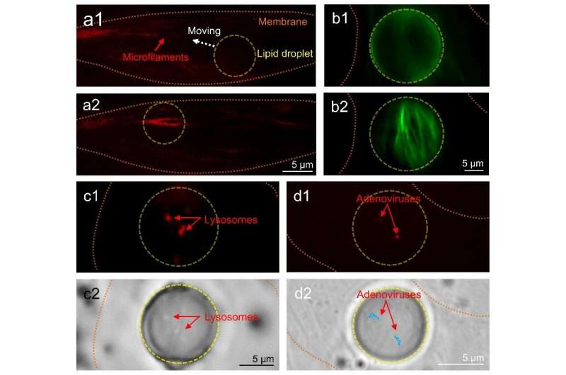
(a) Fluorescence images of the microfilaments (F-actin filaments) of a living adipose cell. The weak fluorescent signals (a1) were efficiently enhanced by the lipid droplet (diameter: 7.7 μm) (a2). (b) Fluorescence images of the microfilaments in a fixed adipose cell with an 18.6-μm lipid droplet. (c) Fluorescence and bright-field images of the lysosomes in a living adipose cell with an 11.3-μm lipid droplet. (d) Fluorescence and bright-field images of the adenoviruses in a living adipose cell with an 8.1-μm lipid droplet, the blue lines represent the movement trajectory of the adenoviruses in 10 min. The fluorescence images were focused on the surfaces of the cells (b1) and the virtual image planes (b2, c1 and d1) formed by the lipid droplets. The bright-field image was focused on the virtual image plane formed by the lipid droplet (c2 and d2). Credit: Xixi Chen, Tianli Wu, Zhiyong Gong, Jinghui Guo, Xiaoshuai Liu, Yao Zhang, Yuchao Li, Pietro Ferraro, and Baojun Li

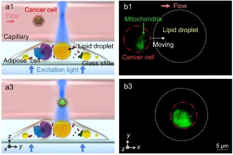
To achieve intracellular imaging of subcellular structures, the lipidic microlenses work in a contact mode in which the collection efficiency of the emitted fluorescent signals is greatly increased by the lipid droplets. As a result, the required excitation power in fluorescence imaging can be reduced by up to 73%. Driven by scanning optical tweezers, the lipid droplets can be moved inside the cell to perform an enhanced intracellular fluorescence imaging of the microfilaments, lysosomes and adenoviruses in living cells. Moreover, due to the refractive index contrast of the lipid droplets to cytoplasm, the long focal length of the lipid droplets can extend the fluorescence enhancement strategies to a non-contact mode that increases the working distance of imaging and detection by the lipidic microlenses. In the non-contact mode, the excitation light is highly converged by the lipid droplets to enhance the fluorescence intensity from the extracellular environment surrounding the cells. In the experiments, an efficient detecting of fluorescent signals of cancer cells in extracellular fluid was accomplished by the lipidic microlenses.

300x250x1
Lipid droplets as endogenous intracellular microlenses
(a) Schematic (side view) of the enhanced detection of cancer cells in fluid by a lipid droplet inside the adipose cell: the lipid droplet acts as a biolens by focusing excitation light into the cancer cell and improving the collection of fluorescent signals at same time. (b) Fluorescence images (top view) of the detecting process. The yellow and red dotted circles indicate the lipid droplet (diameter: 20 μm) and the cancer cell, respectively. The mitochondria of the cancer cell were marked by Mito-Tracker (green). The red arrow indicates the direction of fluid flow in the capillary. Credit: Xixi Chen, Tianli Wu, Zhiyong Gong, Jinghui Guo, Xiaoshuai Liu, Yao Zhang, Yuchao Li, Pietro Ferraro, and Baojun Li

The presented lensing effect of the lipid droplets is expected to find applications with fully biocompatible miniaturized tools for biosensing, endoscopic analysis and single-cell diagnosis. The use of the lipid droplets as intracellular microlenses also provides opportunities to design and construct diverse endogenous photonic devices.


Biophotonic probes for bio-detection and imaging


More information:
Xixi Chen et al, Lipid droplets as endogenous intracellular microlenses, Light: Science & Applications (2021). DOI: 10.1038/s41377-021-00687-3
Provided by
Chinese Academy of Sciences

Citation:
Lipid droplets as endogenous intracellular microlenses (2021, December 27)
retrieved 27 December 2021
from https://phys.org/news/2021-12-lipid-droplets-endogenous-intracellular-microlenses.html

This document is subject to copyright. Apart from any fair dealing for the purpose of private study or research, no
part may be reproduced without the written permission. The content is provided for information purposes only.

Denial of responsibility! NewsConcerns is an automatic aggregator of the all world’s media. In each content, the hyperlink to the primary source is specified. All trademarks belong to their rightful owners, all materials to their authors. If you are the owner of the content and do not want us to publish your materials, please contact us by email – [email protected]. The content will be deleted within 24 hours.

Adblock test (Why?)

728x90x4

Source link

Continue Reading

Science

NASA's Voyager 1 resumes sending engineering updates to Earth – Phys.org

Published

 on


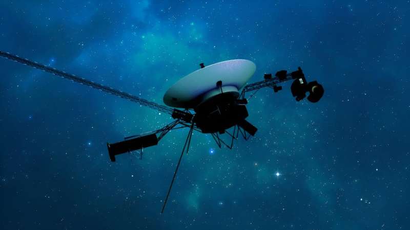
NASA’s Voyager 1 spacecraft is depicted in this artist’s concept traveling through interstellar space, or the space between stars, which it entered in 2012. Credit: NASA/JPL-Caltech

For the first time since November, NASA’s Voyager 1 spacecraft is returning usable data about the health and status of its onboard engineering systems. The next step is to enable the spacecraft to begin returning science data again. The probe and its twin, Voyager 2, are the only spacecraft to ever fly in interstellar space (the space between stars).

Voyager 1 stopped sending readable science and engineering data back to Earth on Nov. 14, 2023, even though mission controllers could tell the was still receiving their commands and otherwise operating normally. In March, the Voyager engineering team at NASA’s Jet Propulsion Laboratory in Southern California confirmed that the issue was tied to one of the spacecraft’s three onboard computers, called the flight data subsystem (FDS). The FDS is responsible for packaging the science and engineering data before it’s sent to Earth.

300x250x1

The team discovered that a responsible for storing a portion of the FDS memory—including some of the FDS computer’s software code—isn’t working. The loss of that code rendered the science and engineering data unusable. Unable to repair the chip, the team decided to place the affected code elsewhere in the FDS memory. But no single location is large enough to hold the section of code in its entirety.

So they devised a plan to divide affected the code into sections and store those sections in different places in the FDS. To make this plan work, they also needed to adjust those code sections to ensure, for example, that they all still function as a whole. Any references to the location of that code in other parts of the FDS memory needed to be updated as well.

NASA’s Voyager 1 resumes sending engineering updates to Earth
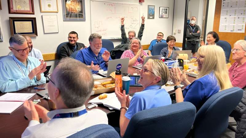
After receiving data about the health and status of Voyager 1 for the first time in five months, members of the Voyager flight team celebrate in a conference room at NASA’s Jet Propulsion Laboratory on April 20. Credit: NASA/JPL-Caltech

The team started by singling out the responsible for packaging the spacecraft’s engineering data. They sent it to its new location in the FDS memory on April 18. A radio signal takes about 22.5 hours to reach Voyager 1, which is over 15 billion miles (24 billion kilometers) from Earth, and another 22.5 hours for a signal to come back to Earth. When the mission flight team heard back from the spacecraft on April 20, they saw that the modification had worked: For the first time in five months, they have been able to check the health and status of the spacecraft.

During the coming weeks, the team will relocate and adjust the other affected portions of the FDS software. These include the portions that will start returning science data.

Voyager 2 continues to operate normally. Launched over 46 years ago, the twin Voyager spacecraft are the longest-running and most distant spacecraft in history. Before the start of their interstellar exploration, both probes flew by Saturn and Jupiter, and Voyager 2 flew by Uranus and Neptune.

Provided by
NASA

Citation:
NASA’s Voyager 1 resumes sending engineering updates to Earth (2024, April 22)
retrieved 22 April 2024
from https://phys.org/news/2024-04-nasa-voyager-resumes-earth.html

This document is subject to copyright. Apart from any fair dealing for the purpose of private study or research, no
part may be reproduced without the written permission. The content is provided for information purposes only.

Adblock test (Why?)

728x90x4

Source link

Continue Reading

Science

Osoyoos commuters invited to celebrate Earth Day with the Leg Day challenge – Oliver/Osoyoos News – Castanet.net

Published

 on


Osoyoos commuters can celebrate Earth Day as the Town joins in on a national commuter challenge known as “Leg Day,” entering a chance to win sustainable transportation prizes.

The challenge, from Earth Day Canada, is to record 10 sustainable commutes taken without a car.

300x250x1

“Cars are one of the biggest contributors to gas emissions in Canada,” reads an Earth Day Canada statement. “That’s why, Earth Day Canada is launching the national Earth Day is Leg Day Challenge.”

So far, over 42.000 people have participated in the Leg Day challenge.

Participants could win an iGo electric bike, public transportation for a year, or a gym membership.

The Town of Osoyoos put out a message Monday promoting joining the national program.

For more information on the Leg Day challenge click here.

Adblock test (Why?)

728x90x4

Source link

Continue Reading

Science

Early bird may dodge verticillium woes in potatoes – Manitobe Co-Operator

Published

 on


Verticillium wilt is a problem for a lot of crops in Manitoba, including canola, sunflowers and alfalfa.

Read Also

Field stress can translate to potato skin flaws.

In potatoes, the fungus Verticillium dahlia is the main cause of potato early die complex. In a 2021 interview with the Co-operator, Mario Tenuta, University of Manitoba soil scientist and main investigator with the Canadian Potato Early Dying Network, suggested the condition can cause yield loss of five to 20 per cent. Other research from the U.S. puts that number as high as 50 per cent.

It also becomes a marketing issue when stunted spuds fall short of processor preferences.

Verticillium in potatoes can significantly reduce yield and, being soil-borne, is difficult to manage.

Preliminary research results suggest earlier planting of risk-prone fields could reduce losses, in part due to colder soil temperatures earlier in the season.

Unlike other potato fungal issues that can be addressed with foliar fungicide, verticillium hides in the soil.

“Commonly we use soil fumigation and that’s very expensive,” said Julie Pasche, plant pathologist with North Dakota State University.

There are options. In 2017, labels expanded for the fungicide Aprovia, Syngenta’s broad-spectrum answer for leaf spots or powdery mildews in various horticulture crops. In-furrow verticillium suppression for potatoes was added to the label.

There has also been interest in biofumigation. Mustard has been tagged as a potential companion crop for potatoes, thanks to its production of glucosinolate and the pathogen- and pest-inhibiting substance isothiocyanate.

Last fall, producers heard that a new, sterile mustard variety specifically designed for biofumigation had been cleared for sale in Canada, although seed supplies for 2024 are expected to be slim. AAC Guard was specifically noted for its effectiveness against verticillium wilt.

Timing is everything

Researchers at NDSU want to study the advantage of natural plant growth patterns.

“What we’d like to look at are other things we can do differently, like verticillium fertility management and water management, as well as some other areas and how they may be affected by planting date,” Pasche said.

The idea is to find a chink in the fungus’s life cycle.

Verticillium infects roots in the spring. From there, it colonizes the plant, moving through the root vascular tissue and into the stem. This is the cause of in-season vegetative wilting, Pasche noted.

As it progresses, plant cells die, leaving behind tell-tale black dots on dead tissue. Magnification of those dots reveals what look like dark bunches of grapes — tiny spheres containing melanized hyphae, a resting form of the fungus called microsclerotia.

The dark colour comes from melanin, the same pigment found in human skin. This pigmentation protects the microsclerotia from ultraviolet light.

Adblock test (Why?)

728x90x4

Source link

Continue Reading

Trending