adplus-dvertising
Connect with us

Health

National Immunization Awareness Week promotes getting caught up

Published

 on


April 24-30 is National Immunization Awareness Week (NIAW) in Canada.

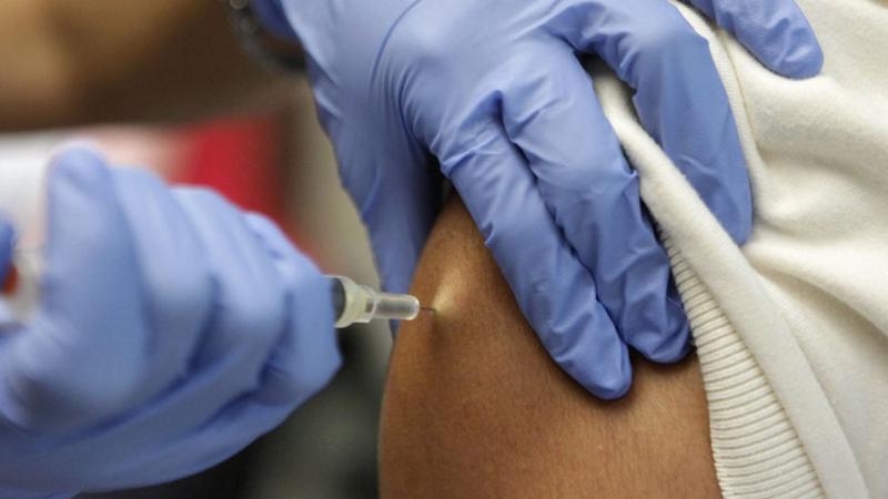
Over the course of the pandemic,Alberta Health Services says, routine immunizations may have been missed or delayed for both children and adults.

300x250x1

Public Health nurses with AHS are currently reaching out to grade 10, 11, 12 families of students who may have missed their routine immunizations due to the pandemic to offer catch up immunizations.

“Today, vaccines continue to contribute to the elimination of diseases, such as measles and cervical cancer. They are also used to help prevent painful conditions like shingles. Most recently, vaccines have played a crucial role in our fight against COVID-19, helping to save the lives of millions of people in Canada. What vaccines have accomplished historically as well as their potential for the future is remarkable,” says Jean-Yves Duclos, federal Minister of Health.


728x90x4

Source link

Continue Reading

Health

Tuberculous monoarthritis of the knee joint – CMAJ

Published

 on


KEY POINTS

  • Joint tuberculosis (TB) is an uncommon manifestation of TB that typically presents with subacute or chronic atraumatic inflammatory symptoms in single, large, weight-bearing joints.

  • In Canada, epidemiological risk factors for TB include extended time spent in countries with high burden of TB, known exposure to TB or being part of a population disproportionately affected by TB (e.g., people born outside of Canada, those of Indigenous ethnicity or those with a history of homelessness or incarceration).

  • Synovial thickening, joint effusions, juxta-articular osteopenia, peripheral osseous erosions and gradual joint space narrowing on magnetic resonance imaging or computed tomography are suggestive of joint TB.

  • Specialized mycobacterial culture is the gold standard for diagnosis of TB, but nucleic acid amplification tests are increasingly accessible and allow for rapid diagnosis of joint TB.

  • Treatment for joint TB should be started promptly for preservation of joint function and prevention of irreversible destruction.

A 47-year-old man presented to the emergency department with 2 weeks of acute-on-chronic pain in his right knee. He described a 5-year history of chronic pain in his right knee, with 1 flare-up 3 years before presentation, which had resolved after a few weeks of ibuprofen use. He had not previously sought care for this problem. He was otherwise healthy, with no chronic medical conditions or previous surgeries, and he tested negative for HIV. He had smoked crystal methamphetamine daily for the last 10 years and had no history of intravenous drug use. He had immigrated from the Philippines 16 years before and had not returned.

When examined, the patient was afebrile with normal vital signs. His right knee was swollen with limited flexion and extension. He did not have any overlying erythema or tenderness to palpation of the bursa or the joint line. He had no other inflamed joints. Initial laboratory results showed a leukocyte count of 8.4 (normal 4–11) × 109/L and an elevated C-reactive protein level of 34.4 (normal < 3.1) mg/L. A radiograph of the patient’s right knee showed bony destruction of the knee joint and an irregular lucency of the medial tibial plateau (Figure 1). A computed tomography (CT) scan showed cortical destruction with peripheral osseous erosions and juxta-articular osteopenia (Figure 2). The patient was admitted to the internal medicine service and the initial treating physicians were concerned that he may have had a malignant disease; they ordered a CT scan of the chest, abdomen and pelvis, which showed mild, biapical lung scarring (Figure 3).

<a href="https://www.cmaj.ca/content/cmaj/195/22/E782/F1.large.jpg?width=800&height=600&carousel=1" title="Radiograph of the right knee joint of a 47-year-old man with joint tuberculosis, showing peripheral osseous erosions (red arrow), irregular lucency of the medial tibial plateau (yellow arrow) and joint space narrowing (white arrow)." class="highwire-fragment fragment-images colorbox-load" rel="gallery-fragment-images-1137217360" data-figure-caption="

300x250x1
Radiograph of the right knee joint of a 47-year-old man with joint tuberculosis, showing peripheral osseous erosions (red arrow), irregular lucency of the medial tibial plateau (yellow arrow) and joint space narrowing (white arrow).

” data-icon-position data-hide-link-title=”0″>Figure 1:

Figure 1:

Radiograph of the right knee joint of a 47-year-old man with joint tuberculosis, showing peripheral osseous erosions (red arrow), irregular lucency of the medial tibial plateau (yellow arrow) and joint space narrowing (white arrow).

<a href="https://www.cmaj.ca/content/cmaj/195/22/E782/F2.large.jpg?width=800&height=600&carousel=1" title="Serial computed tomography scans (coronal view) of the right knee of a 47-year-old man with joint tuberculosis, showing the Phemister triad of tuberculosis arthropathy including juxta-articular osteopenia (yellow arrows), peripheral osseous erosions (red arrows) and joint space narrowing (white arrow, panel A)." class="highwire-fragment fragment-images colorbox-load" rel="gallery-fragment-images-1137217360" data-figure-caption="

Serial computed tomography scans (coronal view) of the right knee of a 47-year-old man with joint tuberculosis, showing the Phemister triad of tuberculosis arthropathy including juxta-articular osteopenia (yellow arrows), peripheral osseous erosions (red arrows) and joint space narrowing (white arrow, panel A).

” data-icon-position data-hide-link-title=”0″>Figure 2:Figure 2:

Figure 2:

Serial computed tomography scans (coronal view) of the right knee of a 47-year-old man with joint tuberculosis, showing the Phemister triad of tuberculosis arthropathy including juxta-articular osteopenia (yellow arrows), peripheral osseous erosions (red arrows) and joint space narrowing (white arrow, panel A).

<a href="https://www.cmaj.ca/content/cmaj/195/22/E782/F3.large.jpg?width=800&height=600&carousel=1" title="Computed tomography scans of the lungs of a 47-year-old man with joint tuberculosis, showing mild biapical scarring (arrows), suspected to be secondary to previous tuberculosis infection. (A) and (B) are serial axial images; (C) and (D) are serial coronal images." class="highwire-fragment fragment-images colorbox-load" rel="gallery-fragment-images-1137217360" data-figure-caption="

Computed tomography scans of the lungs of a 47-year-old man with joint tuberculosis, showing mild biapical scarring (arrows), suspected to be secondary to previous tuberculosis infection. (A) and (B) are serial axial images; (C) and (D) are serial coronal images.

” data-icon-position data-hide-link-title=”0″>Figure 3:Figure 3:

Figure 3:

Computed tomography scans of the lungs of a 47-year-old man with joint tuberculosis, showing mild biapical scarring (arrows), suspected to be secondary to previous tuberculosis infection. (A) and (B) are serial axial images; (C) and (D) are serial coronal images.

Bloody synovial fluid was aspirated from the patient’s right knee joint, with a leukocyte count of 36.7 × 10⁹/L (79% neutrophils, 15% lymphocytes, 6% monomacrophages) and an erythrocyte count of 150 × 109/L, and no crystals or organisms on Gram stain. Cultures for mycobacteria were negative on acid-fast bacilli staining. Given these findings, bacterial septic arthritis was suspected and he was treated with empirical antibiotic therapy (intravenous [IV] ceftriaxone 2 g daily, and IV vancomycin with a target trough of 10–20 mg/L). After 6 days, the patient’s knee had not improved and bacterial cultures from synovial fluid remained negative. The internal medicine team consulted our infectious diseases team.

We suspected tuberculosis (TB) based on 3 factors, namely the chronic nature of the monoarthritis, the pulmonary apical scarring on chest imaging and that the patient had emigrated from the Philippines — designated as a country with a high burden of TB by the World Health Organization (WHO), with the fourth highest national incidence of TB globally.1 We identified no other identifiable risk factors for TB, such as previous incarceration, homelessness or known close contact with someone with TB.

Nucleic acid amplification testing (NAAT) for TB using the GeneXpert MTB/RIF Ultra assay (Cephied) was performed on samples of synovial fluid, which confirmed the presence of Mycobacterium tuberculosis. Smear testing of sputum samples were negative for acid-fast bacilli. We prescribed standard weight-based dosages of rifampin (10 mg/kg/d), isoniazid (5 mg/kg/d) with pyridoxine (25 mg/d), pyrazinamide (25 mg/kg/d) and ethambutol (15 mg/kg/d) and discharged him to continue this therapy under care of the provincial TB program.

Four weeks later, samples of the patient’s synovial fluid showed growth of M. tuberculosis that was susceptible to the first-line anti-TB medications he was receiving. Mycobacterial cultures from sputum samples were negative, suggesting that he did not have pulmonary TB. At follow-up after the 9-month course of treatment, the patient had considerable improvement of his knee pain (pain only with extended use), periarticular and lower leg swelling and range of motion (full flexion and extension). He had returned to work and was ambulating without need for a mobility aid. Given the extensive destruction of the knee joint, we had planned to refer him to an orthopedic surgeon for consideration of knee arthroplasty. However, he was lost to follow-up, and subsequent knee imaging and surgical referral could not be completed.

Discussion

In 2020, the incidence of TB in Canada was 4.7 per 100 000 people, consistent with the previous 10 years.2 However, people born outside of Canada and Canadian-born Indigenous peoples are disproportionately affected.2 Bone and joint TB, including both vertebral and extra-axial joint disease, is an uncommon manifestation of extrapulmonary TB. In Canada, this form of TB accounts for about 2.5% of all TB cases per year, and around half of these cases have vertebral disease.3 In low-income regions, bone and joint TB is more commonly seen among children, usually developing shortly after primary infection. In high-income countries, however, it is most commonly seen among adults, resulting from TB reactivation, as we suspected with our patient.3 Extra-axial disease usually presents as a monoarthritis that affects the knee or hip.

Diagnosis may be difficult and is often delayed by many months, particularly in countries where TB is not endemic.4,5 Key features of TB include the presence of risk factors, the long duration of symptoms and constitutional symptoms. Unlike in bacterial septic arthritis, systemic infectious symptoms such as fevers and chills, rigors or elevated heart rate are usually absent in tuberculous arthritis. Other differential diagnoses include bacterial septic arthritis, crystal arthropathy, systemic rheumatic disease (such as rheumatoid arthritis) and osteoarthritis. Poncet disease, a reactive polyarthritis associated with extra-articular TB disease, should also be considered if several joints are involved and the patient has evidence of nonerosive arthritis.6 Findings from physical examination are nonspecific and include restricted range of motion, swelling and joint effusion.7 Joint erythema, warmth and tenderness may be absent, unlike in crystal arthropathy or bacterial septic arthritis.

Typical radiologic findings in early disease show thickening of the synovium and joint effusions — similar to other causes of monoarthritis such as septic arthritis or crystal arthropathy.8 Early TB arthritis may also be misdiagnosed as osteoarthritis if imaging shows only joint space narrowing.8 Later-stage disease has features of juxta-articular osteopenia, peripheral osseous erosions and joint space narrowing, known as the Phemister triad.9 Some chronic cases may have sinus tract formation.8

Plain radiographs may show the Phemister triad in later-stage disease, but magnetic resonance imaging (MRI) and CT are the best imaging modalities for disease characterization. In particular, MRI offers the most specificity for TB arthritis, but timely availability is often limited; therefore, CT may be used as it offers more specific disease characterization than plain radiographs. 8 However, because joint TB has no pathognomonic imaging findings, particularly early in the disease course, the diagnosis cannot be made based on imaging alone and requires microbiological confirmation.

Diagnosis is typically achieved by synovial fluid analysis or synovial biopsy with microscopy for acid-fast bacilli and mycobacterial culture. However, testing synovial fluid for acid-fast bacilli has a low sensitivity of about 30% and cultures take weeks to detect growth. Mycobacterial cultures also require specialized laboratories that are certified for biosafety containment level 3 and have clinical mycobacteriology accreditation to ensure quality assurance.3 Joint fluid analysis often finds a leukocyte count in the inflammatory range, typically 10–20 × 109/L, but may be higher.7

Newer methods for molecular detection of M. tuberculosis such as NAAT are rapid, more geographically accessible and do not require specialized laboratories. The most studied and widely available NAAT assay recommended by WHO is the automated, cartridge-based GeneXpert MTB/RIF and the newer generation, GeneXpert MTB/RIF Ultra. A recent review of the diagnosis of extrapulmonary TB with the GeneXpert assay found a sensitivity of 60%–100%, depending on specimen type, and a specificity of 87.5%–100% for bone and joint specimens.10 We did not diagnose TB in our patient until 5 years after symptom onset, by which time he had considerable destruction of the knee joint. Despite the presence of joint destruction, TB arthritis typically responds well to medical therapy and is usually treated for 6–12 months. Treatment duration should be extended for patients with increased risk of relapse, such as those with extensive disease at presentation.3 Unlike other causes of septic arthritis, TB arthritis does not require acute surgical management; surgery is reserved for treatment of complications related to joint destruction after completion of TB treatment.3

Conclusion

Clinicians should consider joint TB in patients who present with subacute joint pain and swelling with joint fluid analysis suggestive of an inflammatory arthritis, and in patients who do not respond to standard therapy for inflammatory or septic arthritis. In addition to mycobacterial cultures, NAAT may aid timely diagnosis and management.

The section “Cases” presents brief case reports that convey clear, practical lessons. Preference is given to common presentations of important rare conditions, and important unusual presentations of common problems. Articles start with a case presentation (500 words maximum), and a discussion of the underlying condition follows (1000 words maximum). Visual elements (e.g., tables of the differential diagnosis, clinical features or diagnostic approach) are encouraged. Consent from patients for publication of their story is a necessity. See information for authors at www.cmaj.ca.

Footnotes

  • Competing interests: None declared.

  • This article has been peer reviewed.

  • The authors have obtained patient consent.

  • Contributors: All of the authors contributed to the conception and design of the work. Adrianna Gunton drafted the manuscript, revised it critically for important intellectual content, gave final approval of the version to be published and agreed to be accountable for all aspects of the work.

This is an Open Access article distributed in accordance with the terms of the Creative Commons Attribution (CC BY-NC-ND 4.0) licence, which permits use, distribution and reproduction in any medium, provided that the original publication is properly cited, the use is noncommercial (i.e., research or educational use), and no modifications or adaptations are made. See: https://creativecommons.org/licenses/by-nc-nd/4.0/

Adblock test (Why?)

728x90x4

Source link

Continue Reading

Health

Neighborhood deprivation and depressive symptoms linked to premature aging – News-Medical.Net

Published

 on


Feeling depressed and living in a deprived urban neighborhood could be making you age faster, according to a new study led by researchers at McMaster University.

The findings, published June 5 in The Journals of Gerontology, Series A: Biological Sciences and Medical Sciences, showed that living in urban environments marked by material and social inequities, and having depression symptoms, were independently associated with premature biological aging, even after accounting for individual-level health and behavioral risk factors, such as chronic conditions and poor health behaviors.

Parminder Raina, a professor in the Department of Health Research Methods, Evidence, and Impact at McMaster University, led the research team, which included investigators from the Netherlands, Norway and Switzerland.

300x250x1

Our study used two DNA methylation-based estimators, known as epigenetic clocks, to examine aging at the cellular level and estimate the difference between chronological age and biological age.”

Divya Joshi, study’s first author and research associate in the Department of Health Research Methods, Evidence, and Impact at McMaster

“Our findings showed that neighborhood deprivation and depressive symptoms were positively associated with acceleration of the epigenetic age estimated using the DNAm GrimAge clock. This adds to the growing body of evidence that living in urban areas with higher levels of neighborhood deprivation and having depression symptoms are both associated with premature biological aging.”

Depressive symptoms in the study were measured using a 10-item standardized depression scale. The researchers found an acceleration in the risk of death by one month for every point increase on the depressive symptom score. They theorized that emotional distress caused by depression may result in more biological wear and tear and dysregulation of physiological systems, which in turn could lead to premature aging.

The researchers assessed neighborhood material and social deprivation using two indices that were developed by the Canadian Urban Environmental Health Research Consortium (CANUE) based on 2011 census.

Social deprivation reflects the presence of fewer social resources in the family and community, and material deprivation is an indicator of people’s inability to access goods and conveniences of modern life, such as adequate housing, nutritious food, a car, high-speed internet, or a neighborhood with recreational facilities.

The researchers found an increase in the risk of death by almost one year for those exposed to greater neighborhood deprivation compared to lower neighborhood deprivation.

The study did not find that neighborhood deprivation amplified the effect of depressive symptoms on epigenetic age acceleration.

“Our results showed that the effect of neighborhood deprivation on epigenetic age acceleration was similar regardless of depression symptoms, suggesting that depression influences epigenetic age acceleration through mechanisms unrelated to neighborhood deprivation,” Joshi said.

The research examined epigenetic data from 1,445 participants enrolled in the Canadian Longitudinal Study on Aging (CLSA), a research platform following more than 50,000 participants who were between the ages of 45 to 85 when recruited.

“Longitudinal studies, like the CLSA, are important to confirm associations like those found in this study,” said Raina, the study’s senior author and lead principal investigator of the CLSA.

“By following the same group of participants for 20 years, we will be able to determine whether epigenetic changes are stable or reversible over time. We will also gain insight into the mechanisms that are leading to accelerated epigenetic aging.”

Support for the CLSA is provided by the Government of Canada through the Canadian Institutes of Health Research and the Canada Foundation for Innovation. Additional support for this study was provided by the European Union Horizon 2020 Programme.

Journal reference:

Joshi, D., et al. (2023) Association of Neighborhood Deprivation and Depressive Symptoms With Epigenetic Age Acceleration: Evidence From the Canadian Longitudinal Study on Aging. The Journals of Gerontology Series A. doi.org/10.1093/gerona/glad118.

Adblock test (Why?)

728x90x4

Source link

Continue Reading

Health

Depression, inequity may accelerate aging: study – CTV News

Published

 on


A new Canadian-led study has found that feeling depressed, along with living in a disadvantaged neighbourhood, may lead to premature aging.

The peer-reviewed study, published on Monday in The Journals of Gerontology, Series A: Biological Sciences and Medical Sciences, points to how depression and living in an urban environment – with greater material and social inequities – can influence how a person ages.

Led by researchers at McMaster University in Hamilton, Ont., the study’s authors say this may occur “even after accounting for individual-level health and behavioural risk factors, such as chronic conditions and poor health behaviours.”

300x250x1

“This adds to the growing body of evidence that living in urban areas with higher levels of neighbourhood deprivation and having depression symptoms are both associated with premature biological aging,” team lead Parminder Raina, a professor in the Department of Health Research Methods, Evidence, and Impact at McMaster University, said in a news release.

The researchers used epigenetic data from 1,445 people enrolled in the Canadian Longitudinal Study on Aging, a long-term study that follows about 50,000 participants between the ages of 45 and 85 for at least 20 years.

The study is described as examining how changing biological, medical, psychological, social, lifestyle and economic factors impact a person’s health as they age.

Epigenetics is the study of how behavior and environment can influence how a person’s genes work.

The researchers measured depression symptoms using a standardized 10-item scale.

They say a one-point increase in the score accelerated a person’s risk of death by one month, with the theory being that emotional distress caused by depression could lead to “more biological wear and tear and dysregulation of physiological systems.”

In order to measure “neighbourhood material and social deprivation,” the researchers used a pair of indices, based on the 2011 census, from the Canadian Urban Environmental Health Research Consortium.

They described material deprivation as a person’s inability to obtain resources such as adequate housing, nutritious food, a vehicle, high-speed internet or recreational facilities, while social deprivation refers to family and community connections.

The researchers say greater neighbourhood deprivation increased a person’s “risk of death by almost one year,” but did not worsen the effect that depression had on aging.

In other words, both were independently associated with premature aging.

“Our results showed that the effect of neighbourhood deprivation on epigenetic age acceleration was similar regardless of depression symptoms, suggesting that depression influences epigenetic age acceleration through mechanisms unrelated to neighbourhood deprivation,” Divya Joshi, first author of the study and a research associate at McMaster University, said.

The research team also included members from the Netherlands, Norway and Switzerland.

Adblock test (Why?)

728x90x4

Source link

Continue Reading

Trending